You must be signed in to read the rest of this article.
Registration on CDEWorld is free. Sign up today!
Forgot your password? Click Here!
Advancements in Dentures
Sia Abai, DDS, MMedSc
The patient with terminal dentition or no dentition presents a clinical challenge in the replacement of form and function by the clinician. In particular, the clinician has the ability to replace missing or soon to be missing teeth, along with the supporting bone and soft tissue that has been lost over many years. Etiologic factors contributing to terminal dentition include poor oral hygiene, lack of access to dental care, and educational attainment, and may often be correlated with socioeconomic status.1 Other factors, such as the correlation between systemic and oral health, have been a predictor for tooth loss, resulting in the loss of masticatory function compromising nutritional intake, lower self-perception, and lower quality of life.
Together with the patient, the clinician must overcome the challenges of restoring the form and function of the teeth along with creating harmony in the stomatognathic system of the temporomandibular joints and the muscles of mastication.2 The challenges in denture prosthetics include and are not limited to eating difficulties, soreness of soft tissue, and occlusal errors. A particular challenge in complete dentures is the retention of the mandibular prosthesis.3 Redford et al demonstrated that more than 50% of conventional mandibular complete dentures lack retention and stability.4 Lack of retention and stability is attributed to less surface area and border seal of the mandibular denture compared with the maxillary denture. Additional factors affecting retention and discomfort of the mandibular denture include bone loss, impingement of frenula, loss of depth of vestibule, and inferior alveolar nerve migration with severe atrophy of the mandible.5
Advancements in complete dentures and implant dentistry have led to the enhanced retention of the mandibular denture with implant-retained overdentures, with the dental implants facilitating improved stability and retention of the mandibular complete denture. The concept of implant-retained mandibular overdentures was further legitimized by the McGill Consensus Statement in 2002, establishing the mandibular two-implant overdenture as the first-choice standard of care for edentulous patients.6
Diagnostic criteria for qualification of treatment involve a thorough oral evaluation, panoramic radiograph, and often the use of cone-beam computed tomography (CBCT) scans to evaluate the bone quality and quantity before implant placement. In addition, CBCT scans can reveal vital structures, such as the inferior alveolar nerve, mental foramina, and any anatomical incongruence that can affect location and position of implant placement.7 The implant position is based on the bone volume and must also satisfy esthetic, functional, and biomechanical requirements. A well-fitting final complete mandibular denture that also establishes the esthetics, phonetics, and function of the patient can lead to proper diagnosing, planning, and completion of implant therapy for patients who qualify for this procedure.
Case Report
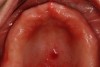
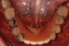
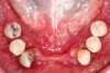
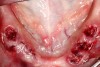
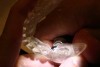
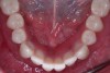
A patient in her mid-50s presented with an ill-fitting maxillary complete denture and a mandibular interim partial denture with six remaining mandibular teeth (Figure 1 and Figure 2). The patient's chief complaint was soreness in the mid-palatal area due to the existing maxillary complete denture and pain in the periodontal supporting structures of the mandibular teeth. A comprehensive evaluation was completed, and several treatment options were presented to the patient. The patient was also referred to and evaluated by an oral surgeon due to the mid-palatal lesion of the hard palate. Pathology was ruled out after a biopsy.
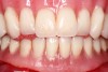
As part of the diagnosis and treatment planning for this case, a CBCT scan was taken and evaluated for bone quality and quantity to present the option of implants for both arches. Because of limited funds and the desire for a solution for the mandibular arch only, the patient opted to have a maxillary complete denture to replace the existing ill-fitting denture, along with an implant-retained mandibular complete denture to address the failing teeth and ill-fitting mandibular interim partial denture (Figure 3).
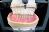
With the treatment plan accepted, the patient was scheduled for preliminary impressions for the fabrication of custom impression trays. The custom impression trays were border-molded at the following appointment to allow the flow of impression material into the depth of the vestibule in order to capture the imperative structures to incorporate into the prosthesis (Figure 4).
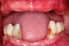
This case was completed with a staged approach because the patient was more concerned about the esthetics of the maxillary arch and existing maxillary complete denture than with the periodontally involved teeth. In lieu of convincing the patient to complete both arches, confidence was built with the patient by addressing her esthetic concerns and continually reaffirming that treatment for the mandibular arch was necessary. The resulting treatment sequence consisted of the fabrication of a maxillary complete denture and a mandibular interim complete denture to allow extraction of the mandibular teeth (Figure 5 and Figure 6).
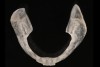
After the extraction of the mandibular teeth and delivery of the complete dentures (Figure 7), the patient underwent a period of 4 to 6 months for the bone graft to heal and for bone deposition in the extraction sites. The mandibular interim denture was evaluated in follow-up appointments after the extractions to provide adjustments and to be relined while the soft tissue and underlying bone healed. The process of fabrication of mandibular complete dentures began after the healing period; a set of complete dentures was fabricated for the patient (Figure 8).
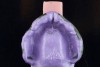
The mandibular final denture served as a guide as to the placement of the mandibular implants to receive an implant-retained overdenture. The process of fabricating a surgical stent involved a duplicate of the final mandibular denture in clear acrylic (Figure 9 and Figure 10), followed by removal of the areas lingual to the incisal and buccal cusps of the teeth. As opposed to a guided surgery surgical template, this surgical guide gave the surgeon an approximation of the location and angulation of implants.
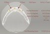
Before the implant surgery, a thorough evaluation of the patient's bone quality and quantity was required. Using a CBCT scan, the evaluation took place with measurements of the allowable space to predictably place the implants (Figure 11). For this case, measurements were taken on an axial slice of the patient's CBCT scan, and the measurements allowed placement of three implants. The method of measurement is to select the correct axial slice, often one that shows both mental foramina. The selection of a mid-point was required. A space of 5 mm anterior to the mental foramina was selected for the distal-most implants. This space allowed the anterior loop of the mental foramina to avoid damage during implant surgery. Although two implants are traditionally treatment planned for the retention of a mandibular overdenture, due to the arch shape of this patient, a third implant was placed at the anterior midpoint of the jaw for more stability.
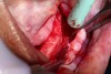
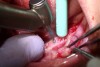
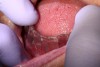
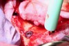
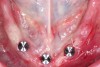
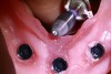
The surgical phase of treatment involved knowledge of anatomy to prevent damage to the inferior alveolar nerve as it exits the mandible bilaterally through the mental foramina and innervates the soft tissue of the anterior segment of the mandible. A full thickness flap was reflected with a releasing incision in the anterior region. The soft tissue was carefully reflected in the area of the mental foramina to prevent nerve damage (Figure 12). With a handpiece and a diamond bur, a mark on the surface of the ridge was placed as a reference point for the location of the mental foramen (Figure 13). This allowed an efficient workflow during the osteotomy. Using the surgical stent and being mindful of the intraoral measurements of the location of the mental foramina, the surgeon proceeded to create the osteotomies in the proper position (Figure 14 and Figure 15).
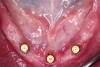
The management of the patient after the implants were placed included education and home care of the existing removable prosthesis. The patient's home care was important because hygiene protocols on cleaning the soft tissue and denture are required to be followed to prevent inflammation through the healing period. After approximately 4 months of healing, the patient's implants were uncovered in preparation for placement of the attachments. The attachments can be placed at the time of second-stage surgery if the implants are uncovered by a minimally invasive procedure. If a full thickness flap across the arch is required, it is recommended that the patient transition from cover screws to healing abutments until the soft tissue heals before moving forward with the delivery of the attachments (Figure 16 through Figure 18).
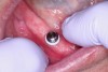
After the attachments were in place, the patient underwent a short period of soft-tissue healing before the pickup of the housing attachments in the intaglio surface of the denture. The attachment was placed on the abutments and the existing final denture was relieved in the areas where the housing attachments were located (Figure 19 and Figure 20).
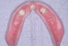
With a hard denture reline material and according to the manufacturer's instructions, the denture's intaglio surface was prepared for the pickup of the housing units chairside. After the housing attachments were picked up, the excess acrylic was trimmed in preparation for delivery of the final implant-retained prosthesis (Figure 21).
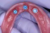
Based on the desired retention of the prosthesis and the particular manufacturer's available options, the black processing units were removed and replaced with the final retention units. In this case, the least retentive retention units were used and demarcated with a blue color (Figure 22).
On final delivery of the implant-retained prosthesis (Figure 23), the patient was given instructions and practiced placement and removal before dismissal. Oral hygiene instructions were given-denture home care is of utmost importance in the longevity of the implants and prosthesis. The patient was instructed to return for follow-up appointments to address any discomfort from soreness from the final prosthesis.
Conclusion
Implant-retained removable prostheses are a predictable solution for patients who require the benefits of improved retention. With proper treatment planning, the surgical and prosthetic portion of this procedure can be predictably delivered to the patient.
About the Author
Sia Abai, DDS, MMedSc
Private Practice
Tustin, California
Lecturer
UCLA Advanced Graduate Prosthodontics
References
1. Latif TM, Vieira AR. Risk factors and comorbidities associated with complete edentulism in individuals younger than fifty years of age. J Dent Oral Health. 2017;4(201):1-6.
2. McCord JF, Grant AA. Identification of complete denture problems: a summary. Br Dent J. 2000;189(3):128-134.
3. Yen HJ, Chen MS, Lin HN, et al. Implant retained overdenture improves the retention and stability by using a Locator System in a mandibular edentulous patient: a case report. J Prosthodont Implantol. 2013;2(2):26-30.
4. Redford M, Drury TF, Kingman A, Brown LJ. Denture use and the technical quality of dental prostheses among persons 18-74 years of age: United States, 1988-1991. J Dent Res. 1996;75(spec no):714-725.
5. Mistry R, Pisulkar SK, Borle AB, et al. Stability in complete dentures: an overview. IOSR J Dent Med Sci. 2018;17(11):36-41.
6. Feine JS, Carlsson GE, Awad MA, et al. McGill consensus statement on overdentures. Mandibular two-implant overdentures as first choice standard of care for edentulous patients. Gerodontology. 2002;19(1):3-4.
7.Scherer MD. Do we need a radiographic guide? A review of the cone-beam computed tomography (CBCT) visualization and treatment planning for narrow-diameter implant overdentures. Implant Practice. 2014;7(3):34-37.